
How to Choose Anterior Fusion Devices in 2026?
Choosing anterior fusion devices in 2026 requires more than comparing product brochures or headline claims. Surgeons must connect device design with patient anatomy, surgical goals, bone quality, and long-term stability. A cage that performs well in a tall, healthy patient may fail to provide the same value in severe osteoporosis. The decision should also consider graft compatibility, subsidence risk, fixation strength, imaging visibility, and revision requirements.
Clinical experience remains important, but it should not replace reliable evidence. Peer-reviewed studies, regulatory status, manufacturer transparency, and post-market safety data can help separate durable performance from attractive marketing. Device selection should reflect the treated level, alignment objectives, neurological condition, and the surgeon’s familiarity with the implantation technique. Small details matter. Endplate preparation, implant footprint, insertion control, and cage positioning can influence outcomes as much as the device label.
There is no universally superior anterior fusion system. That assumption deserves scrutiny. Evidence may be limited for newer materials, especially when follow-up periods remain short. Hospitals should also examine training support, sterilization requirements, inventory reliability, and total procedural cost. These practical factors often shape real-world safety.
A thoughtful 2026 evaluation should combine published research, clinical judgment, patient-specific imaging, and transparent risk discussion. The final choice must serve the patient, not the product category. Even experienced teams should revisit their assumptions when new comparative data appear. In anterior fusion, precision is valuable, but humility is equally necessary.
What Are Anterior Fusion Devices and How Do They Work?
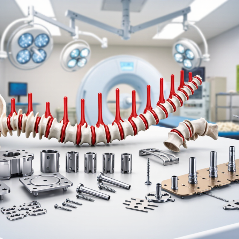
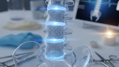
Anterior fusion devices are implants used to stabilize the spine through an approach from the front of the body. Surgeons may use them in the neck or lower back. Common components include an interbody cage, bone graft material, and sometimes a plate with screws.
The procedure usually begins with removing a damaged spinal disc. This can relieve pressure around a nerve or the spinal cord. The surgeon then places a cage between the vertebrae to restore disc height and support alignment. Bone growth through or around the cage helps the neighboring vertebrae join over time. Plates and screws can provide additional stability while fusion develops.
These devices do not move the spine like artificial discs. They aim to create controlled stability. Device selection depends on bone quality, spinal level, alignment, imaging findings, and the patient’s health. A surgeon may also consider the implant’s height, shape, material, and fixation method. Small details matter.
They are not magic.
In practice, a device that fits one patient may be unsuitable for another. Poor positioning, weak bone, smoking, or excessive strain can affect healing. Evidence and surgical experience should guide decisions, but neither replaces a careful examination. Patients should ask how the device supports fusion, what risks apply, and how recovery will be monitored. The plan may need revision when symptoms, scans, or bone healing do not follow expectations.
| Device or Selection Factor | What It Is | How It Works | Potential Advantages | Key Considerations |
|---|---|---|---|---|
| Anterior interbody device | An implant placed in the disc space through an anterior surgical approach. In the lumbar spine, this is commonly used during anterior lumbar interbody fusion (ALIF); anterior interbody devices are also used in cervical procedures. | After disc removal and preparation of the vertebral endplates, the implant helps maintain disc-space height and alignment while bone grows across the treated level. | Can provide a broad area of contact with the vertebral endplates and create space for bone graft material. | The suitable device and approach depend on the spinal level, anatomy, diagnosis, bone quality, and the surgeon’s treatment plan. |
| Cage material: PEEK | Polyether ether ketone, a radiolucent polymer used in some interbody implants. | The cage provides structural support; radiolucency can make it easier to assess the surrounding bone on standard X-rays. | Its imaging characteristics can help clinicians evaluate the fusion area without the cage obscuring it as much as a fully metallic implant may. | Imaging appearance does not by itself establish that fusion has occurred. Material choice should be considered alongside device design and clinical factors. |
| Cage material: porous or solid titanium | Titanium implants may have solid, textured, or porous surfaces, depending on their design. | The implant supports the disc space. Surface features are designed to provide an interface with bone; imaging can show the implant clearly, though metal may affect image appearance. | Available designs can offer different combinations of structural support, surface texture, and imaging characteristics. | Porosity, surface design, and imaging effects vary by device. A material label alone does not predict an individual patient’s result. |
| Integrated fixation | Some interbody devices include screws or other fixation features that attach the implant to adjacent vertebrae. | Integrated fixation is intended to help secure the implant at the treated level. It may be used with or without additional supplemental fixation, as determined by the surgeon. | Can provide a device-specific means of securing the implant without requiring a separate anterior plate in some constructs. | Fixation needs depend on spinal level, stability, bone quality, anatomy, and the overall construct. Not every device or procedure uses integrated fixation. |
| Separate supplemental fixation | Additional instrumentation, such as posterior screws and rods or an anterior plate, used alongside an interbody implant when clinically indicated. | Provides additional stabilization of the treated segment while bone healing and fusion progress. | May be considered when the surgeon determines that the implant alone does not provide the desired stability. | It adds instrumentation and may involve additional surgical steps or an additional approach. The need is patient- and procedure-specific. |
| Implant footprint and height | Footprint describes the implant’s contact area; height describes its dimension within the disc space. | The surgeon selects dimensions intended to fit the prepared disc space and support the vertebral endplates. | A suitable fit can help distribute support across the endplates and maintain the planned disc-space dimensions. | Oversizing or poor fit can create problems, including excessive endplate loading. Dimensions should be selected using patient anatomy and surgical judgment. |
| Lordotic angle and alignment | Some devices are shaped with an angle intended to help restore or maintain a planned spinal alignment. | The implant’s geometry contributes to the position of the vertebrae at the treated level when placed. | May help the surgeon address segmental alignment as part of the overall procedure. | Correction goals depend on the patient’s condition and overall spinal alignment; a device angle alone does not determine the final result. |
| Bone graft material | Bone graft or a graft substitute may be placed within or around the interbody device, according to the device design and surgical plan. | Graft material provides a setting for bone growth across the treated disc space; fusion develops over time rather than immediately after implantation. | Complements the structural role of the implant by supporting the biological process of fusion. | Graft selection, healing, and fusion rates are affected by multiple clinical factors. Follow-up assessment is needed to evaluate healing. |
| Anterior approach and anatomy | The surgeon reaches the spine from the front of the body. Lumbar anterior approaches pass through the abdomen and require consideration of nearby blood vessels and organs. | The approach provides access to the front of the disc space for disc removal, implant placement, and grafting. | Provides direct access to the anterior disc space and can accommodate a comparatively broad implant design in appropriate cases. | Approach-related risks and suitability vary by level and individual anatomy. Preoperative assessment and specialist planning are essential. |
| Choosing a device in 2026 | Compare the intended spinal level, implant dimensions, material, surface design, fixation options, and compatibility with the planned surgical approach. | The device is selected as one component of a treatment plan that also considers diagnosis, bone quality, alignment goals, grafting, and stabilization. | A structured comparison helps match device features to anatomical and surgical requirements. | There is no universally best device. Confirm that the selected device is authorized for its intended use in the relevant jurisdiction and appropriate for the individual patient. |
| Important note | This table provides general educational information, not medical advice or a device recommendation. Anterior fusion devices are selected and implanted by qualified clinicians; patients should discuss individual risks, alternatives, and expected recovery with their care team. | |||
Related Posts
-
2026 Top One Surgical Trends to Watch This Year?
-
2026 How to Choose Lateral Lumbar Interbody Fusion Solutions?
-

Top 10 Disc Fusion Treatments for Global Buyers in 2026?
-
10 Essential Tips for Choosing Surgical Solutions Inc for Your Needs?
-

2026 Top Posterior Cervical Fusion Systems for Global Buyers
-
What is lumbar interbody and why is it important?